
Microscopy in Modern Medicine: What Doctors See That the Naked Eye Cannot
In my clinical practice, one of the most fundamental tools we rely on—yet one that patients rarely think about—is microscopy. Many patients are surprised when I explain that a simple blood or urine sample can reveal detailed information about infections, anemia, kidney disease, and even early signs of more serious conditions when examined under a microscope.
Microscopy allows us to see what the human eye cannot detect, and in many cases, it provides the first clue toward an accurate diagnosis.
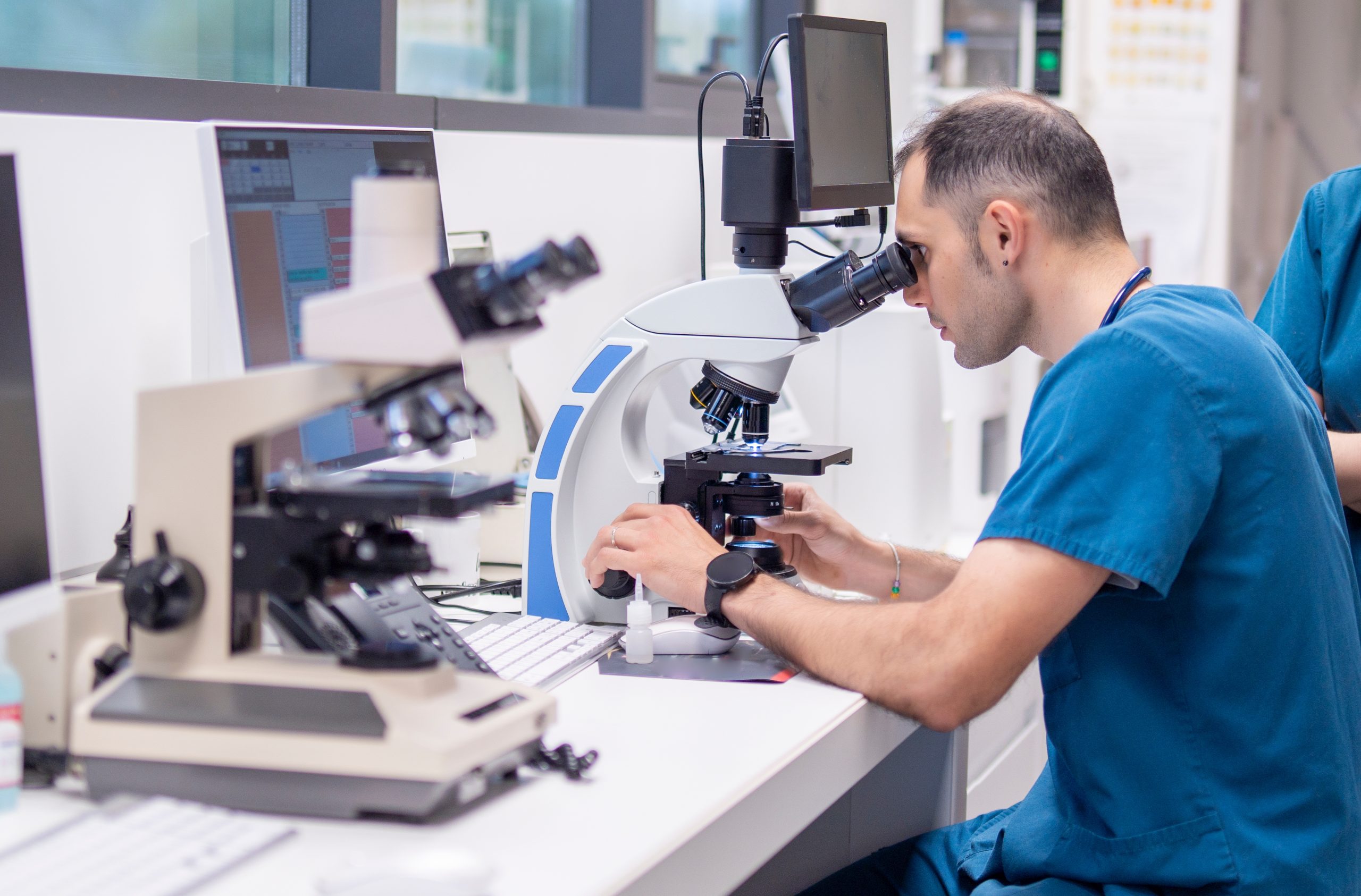
What is Microscopy in Medicine?
Microscopy is the use of a microscope to magnify and examine cells, tissues, and microorganisms. In healthcare, it is commonly used in laboratory diagnostics to evaluate:
- Blood cells
- Bacteria and parasites
- Urine components
- Tissue samples (biopsies)
It remains one of the most reliable and cost-effective diagnostic tools in modern medicine.
How Doctors Use Microscopy in Practice
When I review lab results, microscopy often plays a key role in confirming what I suspect clinically.
For example:
- A patient with fever may have a blood smear examined for malaria parasites or abnormal white blood cells
- A patient with urinary symptoms may have urine microscopy to detect infection or crystals
- A patient with fatigue may have blood cells checked for anemia patterns
Microscopy gives us a direct “visual confirmation” of what is happening inside the body.
Common Types of Microscopy Tests
Blood Smear Examination
Used to assess red and white blood cells, detect infections, and identify blood disorders.
Urine Microscopy
Helps detect urinary tract infections, kidney disease markers, and abnormal substances.
Stool Microscopy
Used to identify parasites or gastrointestinal infections.
Tissue Microscopy (Histopathology)
Performed on biopsy samples to detect inflammation, infection, or abnormal growth.
Why Microscopy is Still Important Today
Even with advanced imaging and automated lab machines, microscopy remains essential because:
- It provides direct visual evidence
- It helps confirm automated lab results
- It allows detection of rare or unexpected abnormalities
- It is highly cost-effective
In many cases, microscopy is the “final confirmation” before a diagnosis is made.
When Patients Should Not Ignore Microscopy Testing
I often recommend microscopy-based tests when patients present with:
- Persistent fever
- Unexplained fatigue
- Painful urination
- Blood in urine or stool
- Suspected infections
These symptoms may appear general, but microscopy helps narrow down the exact cause.
Limitations of Microscopy
While extremely useful, microscopy does have limitations:
- Requires trained laboratory personnel
- Some infections may be too small or few to detect easily
- Results may depend on sample quality
This is why it is often combined with other laboratory tests for accuracy.
Doctor's Insight
From my experience, some of the most important diagnoses begin with something very simple—a slide under a microscope. Patients often expect advanced scans or expensive tests, but many conditions can be identified early through basic microscopy, allowing timely treatment and better outcomes.
– Dr. Balavendrian Anthony
Physician | Palliative Care
International Health
Screening Centre
Evercare Medical Care Centre, Penang, Malaysia.
Frequently Asked Questions
Is microscopy a painful test?
No, microscopy itself is not painful. It is performed on samples like blood, urine, or tissue collected through standard procedures.
Why do doctors need a microscope if I already did a blood test?
Microscopy allows doctors to visually examine cells and detect abnormalities that machines may not fully identify.
How long do microscopy results take?
It depends on the test, but many results are available within a few hours to a day.
Can microscopy detect cancer?
Yes, tissue microscopy (biopsy analysis) can help identify abnormal or cancerous cells.